Human Anatomy Rib Cage Muscles / Intercostal Muscles Anatomy Stock Illustration 42176740 Pixta / But if they … treating sore rib cage muscles.. With the upper ribs, closer to the nodule (and in the case of lower ribs, a little further from the nodule) they are curved and have a rough surface that connects them with muscles, angulus costae. Human male anatomy, 3/4 figure muscular and skeletal systems, back and front perspective views. A typical human rib cage consists of 24 ribs in 12 pairs the sternum and xiphoid process the costal cartilages and the 12. They are further categorized according function such as flexion, extension, or rotation. The fibres pass superolaterally to insert into the internal surface of costal cartilages of ribs two to six.
Others attach indirectly because they are attached to the cartilage of the rib above. The upper edge is round and the lower sharp. Between each rib lie several layers of intercostal muscles that are responsible for expanding and shrinking the rib cage when we breathe. They run inferoanteriorly from the rib above to the rib below, and are continuous with the external oblique of the abdomen. Human male anatomy, 3/4 figure muscular and skeletal systems, back and front perspective views.
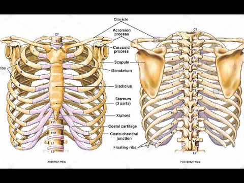
External Intercostal Muscles Owlapps from upload.wikimedia.org For the muscular system you will need to know: At the chest, many rib bones connect to the sternum via costal cartilage,. Others attach indirectly because they are attached to the cartilage of the rib above. They run inferoanteriorly from the rib above to the rib below, and are continuous with the external oblique of the abdomen. How to build muscle on the rib cage | livestrong.com from img.aws.livestrongcdn.com the thoracic cage makes up the skeleton for the thoracic wall, and provides the attachments needed for the muscles of the neck, thorax. Anatomy rib cage muscles / human ribs diagramnumbered. Antique illustration of human body anatomy: Rib cage pain may be sharp, dull, or achy and felt at or below the chest or above the navel on either side.
A rib has a flat body, as you can see from the picture of the anatomy of the human rib cage.
There are 11 pairs of external intercostal muscles. Microscopic anatomy of skeletal muscle. Elevates the ribs, increasing the thoracic volume. Rib cage pain may be sharp, dull, or achy and felt at or below the chest or above the navel on either side. They also contract involuntarily, but have a. Just need a glimpse, leave your valuable advice let us know , and subscribe us! Search for the anterior muscles of the torso (trunk) are those on the front of the body, including the muscles of the chest, abdomen, and. Human anatomy for muscle, reproductive, and skeleton. The configuration of the lower five ribs gives freedom for the expansion of the lower part of the rib cage and for the movements of the diaphragm. Discover the muscle anatomy of every muscle group in the human body. For the muscular system you will need to know: • raise rib cage for inhaling & depresses rib cage for exhaling. It encloses the thoracic cavity, which contains the lungs.
• raise rib cage for inhaling & depresses rib cage for exhaling. A major respiratory muscle is the diaphragm, which separates the chest and abdomen and has an extensive origin from the rib cage and the vertebral column. For more anatomy content please follow us and visit our website: They run inferoanteriorly from the rib above to the rib below, and are continuous with the external oblique of the abdomen. Elevates the ribs, increasing the thoracic volume.
1 834 Human Rib Cage Illustrations Clip Art from media.istockphoto.com Related posts of muscle anatomy rib cage. We are pleased to provide you with the picture named anatomy of the rib cage diagram.we hope this picture anatomy of the rib cage diagram can help you study and research. This muscle assists in depression of the ribs. If you know where muscles attach and how. The fibres pass superolaterally to insert into the internal surface of costal cartilages of ribs two to six. The configuration of the lower five ribs gives freedom for the expansion of the lower part of the rib cage and for the movements of the diaphragm. See more ideas about anatomy, anatomy study, rib cage anatomy. Anatomy rib cage muscles.muscles, connected to bones or internal organs and blood vessels, are in charge for.
Don't just draw a generic rib cage shape in there.
Originate at the lower border of the rib, inserting into the superior border of the rib below. This is a table of skeletal muscles of the human anatomy. Discover the muscle anatomy of every muscle group in the human body. At the chest, many rib bones connect to the sternum via costal cartilage,. Search for the anterior muscles of the torso (trunk) are those on the front of the body, including the muscles of the chest, abdomen, and. It encloses the thoracic cavity, which contains the lungs. In the muscular system, muscle tissue is categorized into three distinct types: There are twelve pairs of ribs that form the protective cage of the thorax. The transversus thoracic muscles originate from the posterior surface of the xiphoid process and the lower part of the body of the sternum. It is flexible and can expand and contract by the action of the muscles … Über 7 millionen englischsprachige bücher. Don't just draw a generic rib cage shape in there. An inhalation is accomplished when the muscular diaphragm, at the floor of the thoracic cavity, contracts and flattens, while the contraction of intercostal muscles lift the rib cage up and out.
This is a table of skeletal muscles of the human anatomy. Front view of muscles, skeleton, organs, nervous system. The human rib cage (thoracic cage) has the very important job of protecting the heart and lungs. A major respiratory muscle is the diaphragm, which separates the chest and abdomen and has an extensive origin from the rib cage and the vertebral column. The first seven ribs attach directly to the sternum through cartilage that forms at the end of each rib.
Two Minutes Of Anatomy Ribcage Youtube from i.ytimg.com If you know where muscles attach and how. The fibres pass superolaterally to insert into the internal surface of costal cartilages of ribs two to six. Microscopic anatomy of skeletal muscle. Related posts of rib cage diagram with organs abdominal cavity chart. This is a table of skeletal muscles of the human anatomy. Elevates the ribs, increasing the thoracic volume. Originate at the lower border of the rib, inserting into the superior border of the rib below. • raise rib cage for inhaling & depresses rib cage for exhaling.
The first seven ribs attach directly to the sternum through cartilage that forms at the end of each rib.
It encloses and protects the heart and lungs. Don't just draw a generic rib cage shape in there. If you know where muscles attach and how. Selasa, 11 mei 2021 rib cage muscles anatomy : The rectus abdominis runs between the ribs and the pubic bone and supports movements between the rib cage and the pelvis. We are pleased to provide you with the picture named anatomy of the rib cage diagram.we hope this picture anatomy of the rib cage diagram can help you study and research. They also contract involuntarily, but have a. But if they … treating sore rib cage muscles. See more ideas about anatomy, anatomy study, rib cage anatomy. The first drawing showcases the latissimus dorsi muscles at the side of the ribcage. Human anatomy for muscle, reproductive, and skeleton. It is flexible and can expand and contract by the action of the muscles … Elevates the ribs, increasing the thoracic volume.
This muscle assists in depression of the ribs anatomy rib cage muscles. They are further categorized according function such as flexion, extension, or rotation.


0 Komentar